Case Summary:
Fibula Free Flap Surgical Guides
Fibula Free Flap Surgical Guides
Patient Diagnosis:
A patient with a history of tumour resection of the left maxilla, reconstructed using a scapular free flap, presented seeking restoration of missing dentition with dental implants.
Given the altered anatomy, variable bone thickness, and unknown implant-bearing capacity of the scapular graft, the clinical team required an objective assessment of feasibility before committing to a surgical pathway.
Planned Procedure/Approach:
The initial intent was to evaluate cone beam scans of the reconstructed maxilla for potential implant placement using virtual surgical planning. The goal was to assess available bone volume, orientation, and implant trajectories within the scapular graft, and—if feasible—progress toward guided implant placement.
During the planning process, concerns arose regarding excessive graft thickness and inconsistent bone stock, leading the surgeons to question whether predictable primary stability could be achieved with implants alone.
What Insight Surgery provided:
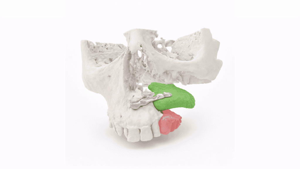
We delivered high-fidelity virtual surgical planning and patient-specific 3D models of the reconstructed maxilla and scapular graft. These allowed the surgical team to interrogate bone thickness, density distribution, and proposed implant positioning in a risk-free digital environment.
Outcome/Benefits:
The modelling directly informed the decision not to proceed with implants at this stage due to the low thickness of the scapula and its ability to accommodate implants, avoiding an unnecessary operative attempt. The virtual model now serves as a shared planning asset for multidisciplinary discussion, with a likely pathway toward bone grafting and a staged return for re-planning and potential guide design for a second-stage procedure.